Project Gallery (6)
Microbial Identification & Morphological Analysis
Overview
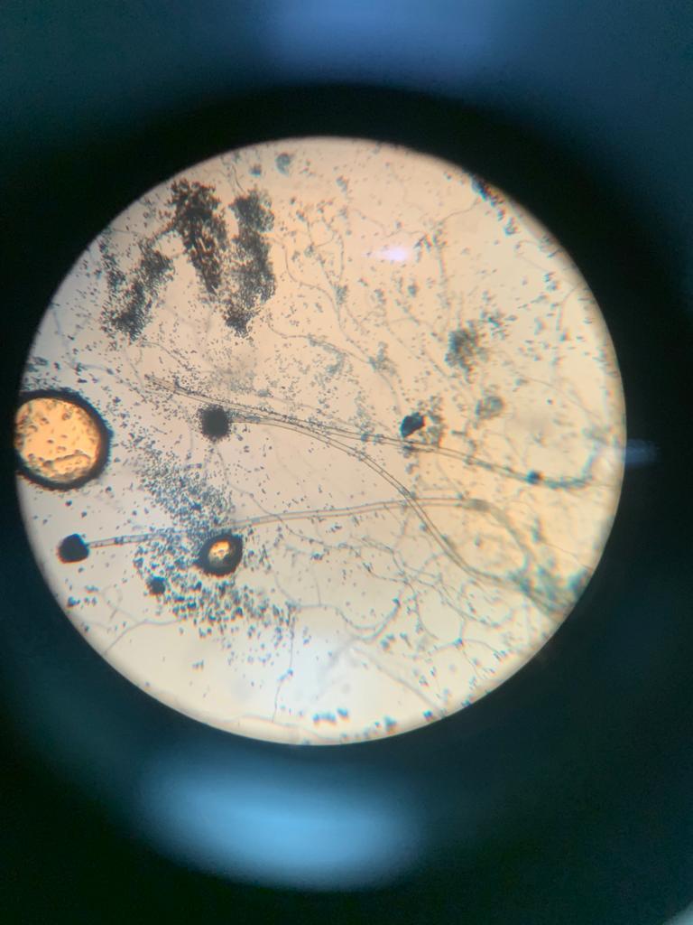
Conducted bacterial and fungal identification through a systematic multi-step approach: differential Gram staining to classify cell wall structure, biochemical tests for species-level identification, slide culture technique to examine fungal colonies, and detailed macroscopic-microscopic morphological observation. All procedures followed established microbiological identification protocols.
The Challenge
Accurate bacterial and fungal identification requires precise execution of multiple sequential techniques — errors in staining timing, reagent concentration, or incubation conditions can lead to misclassification. Colony counting demands consistency and proper dilution technique to obtain countable plates (30-300 colonies).
The Solution
Followed strict timing protocol for each Gram staining step (crystal violet 60s, iodine mordant 60s, decolorizer 10-15s, safranin counterstain 45s). Performed duplicate slides and triplicate plates for result verification. Used proper serial dilution technique for colony counting and recorded all observations using standardized documentation sheets.
Results
Correctly classified bacteria into Gram-positive (purple) and Gram-negative (pink) groups
Identified cell morphology: cocci, bacilli, and arrangements (chains, clusters, diplococci)
Identified fungi macroscopic characteristics: color, texture, radial furrow, growing zone, etc
Successfully correlated macroscopic colony characteristics with microscopic observations
Documented complete identification workflow with photographic evidence